The AIIMS Thursday successfully conducted India’s first craniopagus surgery to separate 28-month-old twins from Odisha, who were joined at the head. AIIMS said Jagga and Kalia, who underwent an 18-hour-long surgery, are “critical” and that they are on ventilator support. A team of 40 doctors from over 12 different super-specialities operated on the twins from Milipada village in Odisha. The twins, who were admitted to the hospital on July 13, were born with an extremely rare condition — joined at the top of their heads, with their bodies at 180 degrees to each other.
Since 1973, only 60 such surgeries have taken place across the world. The last one was reported from Philadelphia in June this year. Out of every 3 million children, only one pair is conjoined; and of all conjoined twins, only 2 per cent are craniopagus. There is a less than 20% chance of survival among craniopagus twins undergoing surgery, data shows.

AIIMS director Dr Randeep Guleria said “post operative care will determine the final outcome” the surgery. “The surgery was a huge challenge as mortality rate was high. With advances in nueroimaging, and with the combined effort of team of specialist, we could successfully separate the twins. There are still on ventilator and are gradually coming out of the anaesthesia. The next few days will be critical. We are observing them very closely. The next journey will continue to be stormy. There is no skull, instead there is a skin graft. The post-operative care will determine the final outcome of the surgery,” Dr Guleria said.
Professor A K Mahapatra, chief of the neurosciences centre at AIIMS, who headed the team, said “the blood pressure and heart rate of the twins are stable”. “In this stage, we faced challenges of blood loss and there was massive blood transfusion,” he said.
In this case, 70% of the brain was infused in one twin, and the rest 30% was infused in the other. They shared a common blood supply channel to the brain. In the first stage, a new venous bypass created. “After that we waited for the alternative pathway to develop and to build up the nutrition and blood level. Then we decided to go for the separation,” Dr Mahapatra added. Professor S S Kale, head, neurosurgery dept, said the “next 18 days” will be critical to decide the outcome of the surgery.
Challenges the doctors had to face
Survival rate differ: one has weak heart; the other weak kidney
The survival rate depends, according to the specialists, on the functioning of key organs. Jagga and Kalia, had blood circulation that was common to “certain extent”, due to the fusion near the vertex, the upper surface of the head. Initially, Jagga was considered healthy and Kalia weak. ““What we used to give to Kalia to treat his infection would go into Jagga’s blood stream. Hence, Kalia was weaker than Jagga,” says Dr Mahapatra. However, after the separation, doctors say that Jagga’s heart is weak and Kalia’s kidney is under pressure. “Now it is a 180 shift. Jagga is weaker than Kalia due to a weak heart. The volume of blood entering Jagga’s heart is more, due to which his heart has to pump more blood. This has made his heart weak. Kalia, is getting lesser fluid, which is resulting in lesser urine output. This will affect kidney functioning,” Dr Guleria said.
Long duration of anesthesia
Story continues below this ad
Professor Girija Prasad Rath from Department of Neuro Anaesthesia said that “long duration” of the surgery was the biggest challenge. The other complication for team included managing “messy blood clots and entry of outside air into circulation”. “We had to make two separate teams of anaesthetists. Both teams had to work with extreme co-ordination. Any small lapse could have resulted in death. There was massive fluid shift. This had to be replaced. The other big challenge was to maintain electrolyte balance in the body. Another challenge was the possibility of hypothermia, that occurs when your body loses heat faster than it can produce heat,” Rath said.
Reconstruction of skin
The skin, skull and brain were shared. And reconstruction of skin to protect the brain from infection was considered the most crucial aspect of the surgery.
“Reconstruction of skin is very important. Children have a big head compared to adults. It is fifteen percent of the body. The other challenge is that in this case brain was uneven, the demand of skin was high. We first expanded the parent skin. There was swelling in brain and we fell short of the skin. We took skin from back and thigh. Then we reconstructed and covered the brain,” Professor Maneesh Singhal, Department of Plastic Surgerysaid.
Neurology and imaging
The nuero imaging team, that captures the images of the nervous system says that even capturing high three-dimensional images of brain structures, using MRI was a challenge. “There were two heads that were attached. We had the area of interest and then increase the size of the image so that we can see both the heads at one time. This was a challenge,” said a specialist.
Story continues below this ad
The pediatric neurology team says that Kalia is now suffering from seizures, which is a challenge. “Both of them did not have seizure to begin with. However, after surgery Kalia had refractive seizures,” says the specialist. Refractive seizures occur when medicines fail to bring seizures under control. “They had a common venous system. We gave Kalia the doze, but half of it went to the other kid. This was a challenge,” says the specialist.

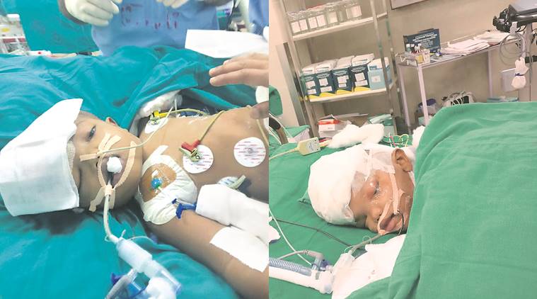 Kalia (left) and Jagga, after the surgery. (Express Photo)