


© The Indian Express Pvt Ltd
Tags:
Researchers at the University of Oldenburg are using electron microscopy images of the coronavirus SARS-CoV-2 to generate images that provide, for the first time, a highly detailed impression of the infection process. The new method relies on machine learning.
As the University says on its website, these days “every child knows what a coronavirus looks like: something like a curled-up hedgehog with spikes that are wider at the top than at their base”. However, many of the images in circulation are illustrations, while an electronic microscope is needed to obtain a direct image of the virus itself. However, “images of SARS-CoV-2 taken with these devices are quite blurred and appear flat or 2-dimensional,” the University quoted researcher Jörg Lücke as saying.

Now the team has made its first major advances. The team has published its preliminary results on a preprint server (not yet peer-reviewed). The researchers are currently conducting further tests, and plan to present their work at an international conference in the near future.
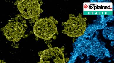
One of the images shows SARS-CoV-2 viruses infecting a cell. In the colourised image, the pathogens look like spherical objects with irregularly shaped protrusions (spike proteins). “However, they are not spiky, but rather rounded in shape, resembling little trees that lean in different directions,” Lücke was quoted as saying. The fact that these tiny structures are recognisable that shows an entire infection scene testifies to how detailed the new images are, he said.
Source: University of Oldenburg


